Insights into all the latest

Research in the News

Tracking the pulse of the latest spinal cord injury research news — from lab breakthroughs to clinical milestones.

Author's Current Top 3 to Read

1

31 JUL 2026

Breathing After High Cervical SCI Without a Ventilator: The Surgery More People Should Know Exists
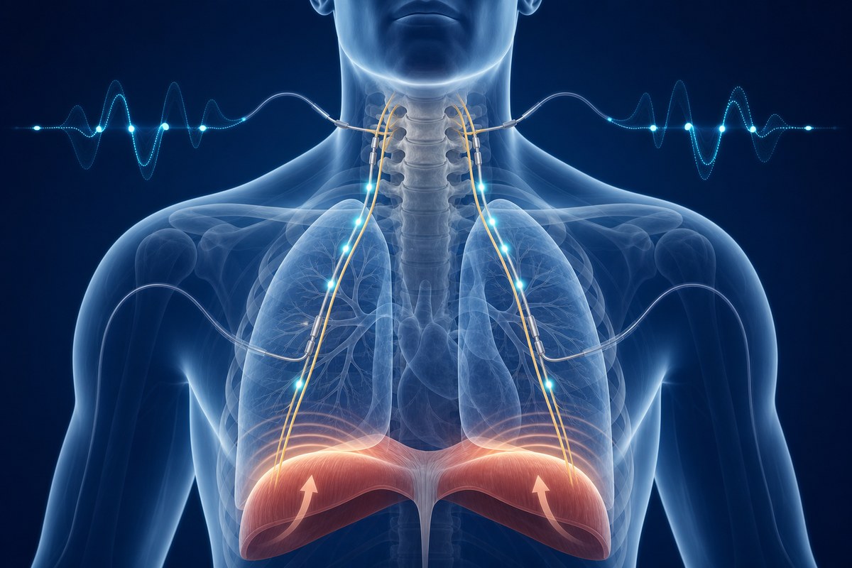
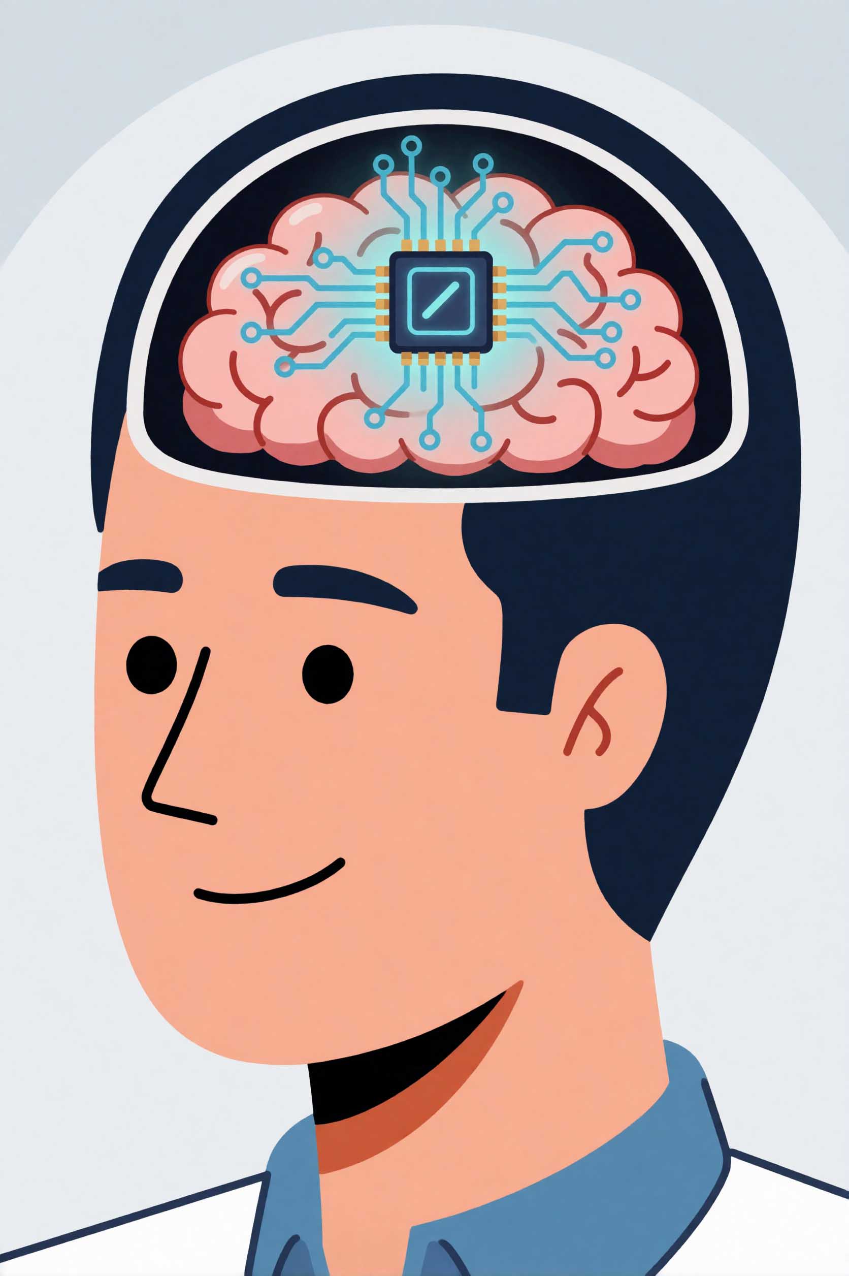
When two-year-old Maverick sustained a high cervical spinal cord injury in a crash, his parents were told he would never breathe without a machine. In January 2026, surgeons at Oklahoma Children's made him the youngest person in the world to undergo phrenic nerve stimulation — a procedure that bypasses the injured cord to restore breathing. Here's what it is, who it can help, and why it isn't more widely known.

Deep Dive

Most people with a high cervical spinal cord injury spend the rest of their lives on a ventilator. Not because their lungs have failed. Not because their diaphragm has stopped working. But because the signal the brain sends to start breathing — travelling down through the spinal cord to the phrenic nerve, the nerve that controls the diaphragm — can no longer get through.

That signal pathway is broken at the cord. But the phrenic nerve itself, in most high cervical injuries, is completely intact. And that is the opening that phrenic nerve stimulation — also called diaphragmatic pacing — exploits.

On 30 July 2026, Oklahoma Children's OU Health published the story of Maverick, a Missouri toddler who suffered a catastrophic cervical SCI in a road crash at 18 months old. He became the youngest person in the world to have phrenic nerve stimulators implanted following a spinal cord injury — at just over two years old, in January 2026. He is now breathing with his own diaphragm, electrically triggered, without a ventilator for stretches of time and progressing further.

His family says they wish more people knew this procedure existed. They're right to say so.

Why breathing fails after high cervical SCI

The phrenic nerves originate in the cervical spinal cord at the C3, C4, and C5 levels — roughly mid-neck. They travel down through the chest and connect to each side of the diaphragm, the dome-shaped muscle that does most of the work of breathing. When the brain signals a breath, the message travels down the cord, exits at C3–C5, and drives the diaphragm to contract and pull air into the lungs.

A spinal cord injury above C3 severs this chain at the cord. The phrenic nerves below the injury remain intact. The diaphragm remains intact. There is simply no signal reaching either of them.

This is where the ventilator steps in: it pushes air into the lungs mechanically, doing the work the diaphragm can no longer do. It is life-saving. But it has real downsides — which is exactly why phrenic nerve stimulation exists.

What phrenic nerve stimulation is

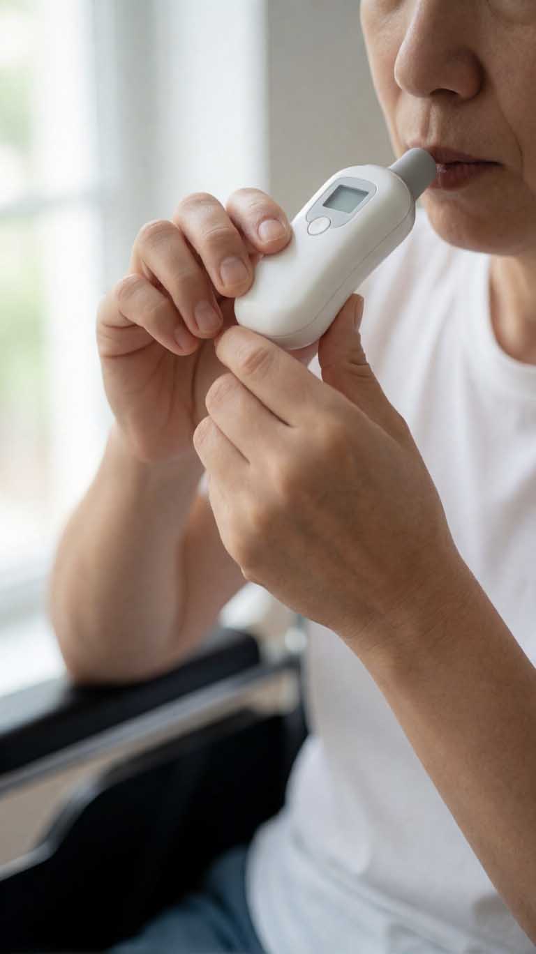
The procedure places small electrodes surgically around or near the phrenic nerve (or at the nerve's entry points into the diaphragm, depending on approach). These electrodes connect to a receiver implanted under the skin. An external control unit transmits electrical pulses through the skin to the receiver, which passes them on to the electrodes, which cause the phrenic nerve to fire — and the diaphragm to contract. A breath follows.

The timing and intensity of those pulses are adjusted to produce a normal breathing rhythm. With conditioning over weeks to months — the diaphragm is a muscle, and needs to be rebuilt after disuse — many patients go from a few hours off the ventilator each day to spending most or all of the day breathing with the pacer. Some transition off the ventilator entirely.

There are two main device types. The conventional approach uses an electrode on the phrenic nerve connected to a subcutaneous receiver and an external radiofrequency transmitter worn on the skin. The Diaphragmatic Pacing System (DPS) places four electrodes directly into the diaphragm at the nerve insertion points, connected to an external pulse generator through a socket at the skin surface. Each approach has clinical uses; the DPS in particular allows stimulation even when the phrenic nerve higher up is partially compromised.

Who qualifies

The key requirement is that the phrenic nerve itself must be intact and functional. Before surgery, phrenic nerve conduction studies — electrical tests of nerve function — confirm whether the nerve can carry a signal.

Patients with SCI above C3 are the clearest candidates: the injury is above where the phrenic roots exit the cord, so the nerve below is undamaged and can be stimulated at the neck, chest, or diaphragm.

Mid-cervical injuries (at C3–C5 itself) are more complex, since the injury may directly involve the nerve roots. In these cases, direct diaphragmatic pacing through the DPS system can sometimes still work by stimulating the nerve at the diaphragm rather than higher up.

Beyond SCI, the procedure is used in congenital central alveolar hypoventilation syndrome (where the brain's breathing control fails), brainstem tumours, Arnold-Chiari malformations, and certain neuromuscular conditions.

How the surgery is done

Surgeons can reach the phrenic nerve three ways. The cervical approach accesses the nerve directly in the neck — the most direct route, typically used for high SCI. The thoracic approach uses keyhole video-assisted chest surgery (VATS) to reach the nerve as it runs alongside the heart. The diaphragmatic approach is laparoscopic — small incisions in the abdomen — and places electrodes where the nerve meets the diaphragm. This is the route used for the DPS device and is available even when the nerve higher up is unreliable.

Both sides are usually done two weeks apart to allow recovery between procedures.

Why it beats a ventilator for most patients

Ventilators save lives, and many people with high SCI live full lives on them. But the practical burden is real: a ventilator requires a tracheostomy (a permanent opening in the windpipe), carries ongoing infection risk, needs an uninterrupted power supply, and is a disconnection risk. Speaking is harder. Taste diminishes over time. Mobility is constrained.

Phrenic nerve stimulators are a closed system once implanted. Most patients keep their tracheostomy initially as a safety backup, but the breathing itself comes through the pacer rather than the machine. Speech follows the natural rhythm of the pacer. Taste is preserved. The device is small and unobtrusive. Because the diaphragm contracts naturally — even if electrically triggered — breathing mechanics are closer to normal, which is better for long-term lung health.

In February 2026, NICE in the UK issued guidance (HTG727) formally endorsing phrenic nerve pacing for ventilator-dependent patients with high cervical SCI — a signal that clinical consensus around the technology is consolidating.

Why it isn't more widely known

Phrenic nerve stimulation has existed since the 1970s — Christopher Reeve used a diaphragmatic pacer in his later years. The technology has improved significantly: devices are smaller, laparoscopic implantation is less invasive than early open surgery, and FDA approval has made the DPS accessible across the US.

But it remains a specialist procedure, concentrated at a small number of centres with specific experience. Awareness among patients, families, and even some clinicians is still limited — which means people who might benefit are sometimes not told it exists. Maverick's case, as the youngest patient in the world to undergo the procedure after SCI, brings it to a wider audience at a moment when guidance and availability are both moving in the right direction.

Reader Q&A

Does everyone with a high cervical SCI qualify?

Not automatically — phrenic nerve function has to be confirmed first through nerve conduction tests. Most people with injuries above C3 do qualify, since the phrenic nerve below the injury is typically intact. People with injuries at C3–C5 need careful assessment, but some still qualify via the diaphragmatic approach. A specialist centre with experience in the procedure is the right place to be evaluated.

Can it replace the ventilator completely?

For many patients, yes, at least for daytime use. Some manage without the ventilator at all. The process involves a conditioning phase — the diaphragm has usually weakened from disuse and needs time to rebuild — which takes weeks to months. Others use the pacer during the day and non-invasive ventilation at night. The combination depends on the individual. Tracheostomies are often kept in place initially as backup, and removed later once breathing is stable.

What age can it be used from?

Maverick's surgery extends the known lower age limit to just over two years old. The Christopher & Dana Reeve Foundation notes that paediatric use of diaphragmatic pacing has actually driven adoption faster than in adults, partly because children's developing nervous systems are more adaptable. Surgeons experienced in paediatric implantation are needed for young patients — but the procedure is not age-restricted in principle.

Is it available in the UK?

Yes, at specialist centres, and with growing clinical support. The February 2026 NICE guidance (HTG727) formalises it as a recommended option for ventilator-dependent high cervical SCI patients, which should strengthen access through NHS pathways. Referral to a specialist spinal or respiratory centre is the starting point.
Read full article → (external link)
2

15 JUL 2026

How to train your breathing after SCI: intensity beats everything else

A study of 81 people with SCI at Switzerland's leading rehabilitation centre found that training intensity and duration predict respiratory improvement far more than injury level or personal characteristics.

Deep Dive

If you have a spinal cord injury — particularly at a cervical or high thoracic level — the muscles that control your breathing are likely affected. Most people with SCI know this in a general way: reduced lung capacity, difficulty clearing the chest after illness, or shortness of breath with effort. What's been less clear is which training approach actually improves this, and whether the severity of your injury limits what's possible.

New research from the Swiss Paraplegic Centre in Nottwil — one of Europe's leading SCI rehabilitation hospitals — gives a more concrete answer than we've had before. The headline finding is both simple and actionable: train hard, train often, and keep going for at least three months. Almost everything else is secondary.

What the study did

Researchers analysed data from 81 people with SCI who completed respiratory muscle training programmes at the Swiss Paraplegic Centre. Participants spanned a range of injury levels and severities — both complete and incomplete injuries — as well as different ages, body types, and baseline fitness levels. This mix was deliberate: the researchers wanted to understand which factors predicted better respiratory outcomes at the end of training.

The training used inspiratory muscle training (IMT) devices — small handheld tools that add resistance to your breathing, similar in principle to a breathing weight. Participants trained at varying intensities and for varying durations as part of their rehabilitation plans.

What they found

When the researchers modelled which factors predicted improvement in respiratory muscle strength, training intensity and programme duration emerged as the dominant predictors — by a significant margin. Personal characteristics like age, sex, and body weight had comparatively little predictive power. Injury characteristics — level, AIS classification (complete vs incomplete), and time since injury — also mattered far less than the training variables themselves.

The practical implication is striking: someone with a complete C4 injury who trains consistently at high intensity for four months is likely to see more improvement than someone with an incomplete C6 injury who trains at moderate intensity for six weeks. The injury doesn't cap your gains as much as the training approach defines them.

The researchers' specific recommendations: train at the highest intensity you can sustain, three to five times per week, for a minimum of three months.

Why respiratory muscle strength matters so much

Pneumonia is the leading cause of death in people with AIS A (complete motor and sensory loss) and AIS B (complete motor loss, some sensory preserved) spinal cord injuries. The primary reason is that impaired respiratory muscles make it harder to cough forcefully and clear the airways of secretions — a problem that becomes critical during a chest infection.

Stronger respiratory muscles translate directly into a more effective cough, better ability to manage respiratory illness at home, and — critically — a reduced risk of pneumonia requiring hospitalisation or mechanical ventilation. The stakes are high enough that this is one area of rehabilitation where the evidence really matters.

Why this study is worth paying attention to

The Swiss Paraplegic Centre treats around 300 new SCI patients a year and carries one of the largest longitudinal SCI rehabilitation datasets in the world. Research from this centre tends to be grounded in clinical reality rather than tightly controlled experimental conditions — these were real patients doing real rehabilitation, not a highly selected research cohort. That makes the findings more directly applicable to practice.

The study also addresses a question that's been genuinely uncertain: does it matter who you are, or does it matter what you do? The answer, clearly, is the latter. That's useful information for patients deciding whether to invest in a training programme, and for clinicians deciding how to structure one.

Reader Q&A

Should I be doing respiratory muscle training? If you have a cervical or upper thoracic SCI and you ever notice difficulty clearing your chest, shortness of breath, or recurring respiratory illness, then yes — it's worth discussing with your physiotherapist or rehabilitation doctor. The evidence is now reasonably strong that structured training helps, and the risk is low.

What kind of device is used? Inspiratory muscle training (IMT) devices are small handheld tools you breathe through against adjustable resistance. They're available without prescription and relatively inexpensive. Your physiotherapist can advise on the right resistance level and technique for your situation.

Does this apply to incomplete injuries too? Yes. The study included both complete and incomplete injuries, and the key finding — that training intensity matters more than injury characteristics — applied across the range.

What if I've had my SCI for years? Is it too late to benefit? Time since injury was not a strong predictor of training response in this study, which suggests it's never too late to benefit. That said, starting respiratory training earlier in rehabilitation tends to establish better habits and baselines — so earlier is generally better, but later is still worth doing.

Read full article → (external link)
3

15 JUL 2026

China performs first commercial brain-computer interface surgery for spinal cord injury

On July 13, a 35-year-old man with cervical SCI became the first person in the world to receive a commercially approved implanted brain-computer interface, during a six-hour surgery at Huashan Hospital in Shanghai.

Deep Dive

Last Sunday, a surgical team at Huashan Hospital — one of China's most prominent neurosurgical centres, affiliated with Fudan University in Shanghai — spent six hours implanting a small array of electrodes into the brain of a 35-year-old man who had lost voluntary use of his hands following a cervical spinal cord injury.

This wasn't an experimental procedure in the usual sense. The device they used — the NEO system, developed by Chinese company Neuracle Technology — had just received commercial approval from China's National Medical Products Administration (NMPA), the country's equivalent of the FDA. This surgery was the first performed under that approval.

If that sounds significant, it is. Until last week, no implantable brain-computer interface had ever been granted commercial approval anywhere in the world. Every previous BCI implanted in a person with paralysis — including those in US Neuralink trials — was done under research exemptions, not as a licensed product a doctor could simply prescribe. We covered the regulatory milestone when it was announced earlier this week; Sunday's surgery is the next chapter.

What the device does

The NEO system is designed for people with cervical SCI — specifically those with injuries between C3 and C7 — who have lost voluntary control of their hands and fingers. The device consists of electrodes implanted into the motor cortex, the part of the brain that controls voluntary movement. It reads the electrical signals the brain generates when the person *thinks* about moving their hand, and translates those signals into commands that can stimulate muscles or control external devices.
The goal is what researchers call "thought-controlled muscle rehabilitation" — restoring functional hand use by creating a new pathway between brain and muscle that bypasses the damaged cord.

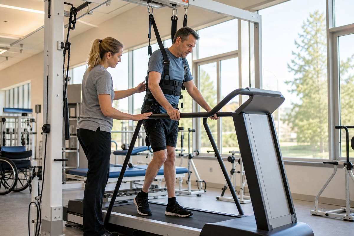
Following the surgery, the patient will need to commit to six hours of rehabilitation per day to train the system and rebuild functional connections. That's intensive, and it underscores that this isn't a passive implant — it requires significant sustained effort from the person receiving it.
Why China, and why now

China has an estimated 3.7 million people living with spinal cord injuries — one of the largest SCI populations in the world. Neuracle Technology has been developing the NEO system for several years, with clinical trials demonstrating safety and some functional benefit sufficient for the NMPA to grant commercial approval.
The specific evidence package used to support the approval hasn't been published in full, but the fact that a sovereign national regulator — with its own evidence standards — granted commercial status to an implantable BCI is notable. No other country has done this yet.

What this doesn't mean yet

It would be easy to read "commercially approved BCI" and assume this technology is now broadly available. It isn't. The NEO system is approved in China. It has not been reviewed by the FDA, the European Medicines Agency, or the MHRA. The approval pathway in China, while rigorous, differs from Western jurisdictions, and each regulator will require its own evidence review.

Access will also depend on cost, surgical infrastructure, and — critically — post-surgical rehabilitation resources. Six hours of therapy daily is a serious commitment, one that requires substantial clinical support to sustain.

None of this diminishes what happened on Sunday. For the first time in history, an implantable BCI for paralysis has moved from experimental to commercial, and the first patient has received it. The regulatory door has been opened. The scientific and commercial pressure to bring equivalent devices through the FDA and European processes will only grow.

Reader Q&A

Could someone with SCI get this surgery right now? Not outside China, and even within China, access will depend on cost, clinical eligibility, and availability of surgical centres experienced with the procedure. This is a brand-new commercial product — the first surgery was performed last Sunday.

Is the NEO system the same as Neuralink? No. Neuralink is a US company with its own design still in clinical trials. Neuracle Technology is a separate Chinese company whose device has now received commercial approval — a stage Neuralink has not yet reached.

Does this mean there's a cure for hand paralysis after SCI? No. The NEO system creates a new communication pathway between brain and muscles, but it doesn't repair the spinal cord. It's an assistive technology that, with intensive rehabilitation, may allow some people to regain functional hand use. The extent of recovery will vary by individual.

When might something like this be available in the UK, US, or Europe? Genuinely unknown. It would require separate regulatory submissions, clinical trials acceptable to the FDA or MHRA, and likely several more years of development. The China approval is a significant milestone, but it doesn't shorten any other regulator's pathway.
Read full article → (external link)

Latest SCI Developments

Filter by main category

Research Regenerative Medicine 17 SEPT 2026

Why nerve fibres don't grow back: a plain-English map of the obstacles

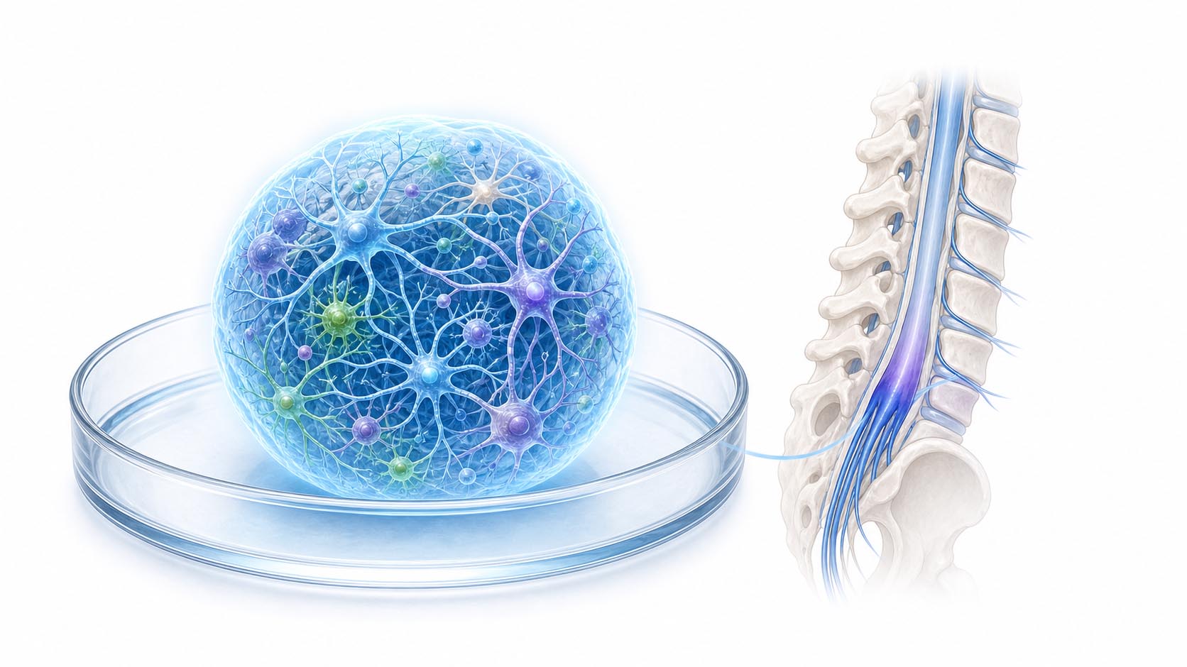
A review in CNS Neuroscience & Therapeutics lays out both reasons axons fail to regrow after spinal cord injury — the hostile environment around them, and the fact that adult neurons have switched off their own growth programme — and why every strategy aimed at one of them keeps stalling on the other.

Read More

Deep Dive

If you have followed spinal cord research for any length of time, you have read a lot of headlines about promising results in mice that never became treatments. A review published in *CNS Neuroscience & Therapeutics* in July 2026 by Jiaxin Gao and colleagues is useful precisely because it explains why that keeps happening.

Note the date: this is a July paper that resurfaced in this week's alerts, not new work. But as a map of where the field actually is, it holds up.

Two problems, not one

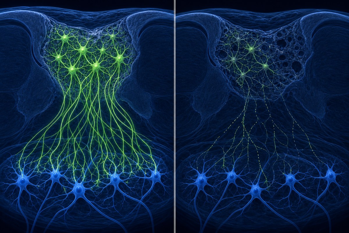
Functional recovery after spinal cord injury depends on axons — the long fibres neurons use to carry signals — regrowing across the damaged region and reconnecting. They almost never do. The review's organising point is that there are two independent reasons for this, and they need different solutions.

The extrinsic problem: the neighbourhood turns hostile.

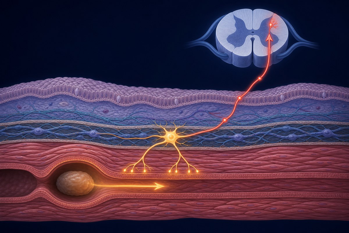
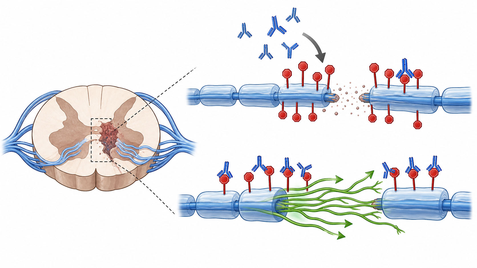
After injury, inflammation and glial activation reshape the damaged area into a glial scar — a dense boundary of cells and matrix around the lesion. Early on the scar is doing useful work: it walls off the inflammation and protects tissue that survived. Left in place for months, though, it becomes both a physical wall and a chemical one.

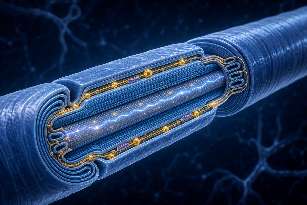
Two families of molecules do most of the chemical blocking. Chondroitin sulfate proteoglycans (CSPGs) are components of the extracellular matrix — the scaffolding between cells — that accumulate in the scar. Myelin-associated inhibitors are released from the debris of damaged myelin, the insulating sheath around nerve fibres. Both switch on signalling inside the growing axon that causes its growth cone — the exploratory tip that finds the way forward — to collapse. The fibre doesn't get blocked so much as told to stop.

The intrinsic problem: the neuron has stopped trying.

During development, neurons run an aggressive growth programme. As they mature, they shut it down — sensibly, since you do not want the wiring in an adult brain rearranging itself. The consequence is that an adult neuron, even placed in a perfectly permissive environment, has limited capacity to regrow a long axon. The instructions are switched off.

Why this explains the graveyard of promising results

Clear the scar, and the neurons still lack the drive to grow. Reactivate the growth programme, and the fibres run into the chemical wall. Either approach alone produces a modest effect in a rodent, gets published, and stops there.

The review catalogues the strategies being pursued against both problems: modulating glial scar dynamics rather than simply removing it, degrading inhibitory matrix components (chondroitinase ABC, an enzyme that chews up CSPGs, is the long-standing example), rebuilding a growth-permissive environment, stem cell transplantation, biomaterial scaffolds that give fibres something to grow along, switching intrinsic growth programmes back on, and neuromodulation — using electrical stimulation to drive activity in surviving circuits.

The authors' conclusion is that meaningful repair will require combinations of these, targeting several mechanisms at once.

What's actually blocking translation

This is the part most worth reading. The review names five specific barriers between a working rodent experiment and a human therapy:

Timing. Many interventions only work in a narrow window after injury. Most people with SCI are years past that window.

Delivery. Getting a biological agent to the right place inside the spinal cord, at the right concentration, without surgery that causes its own damage, is unsolved for most candidates.

Biosafety. Reactivating growth programmes in adult neurons means reactivating machinery the body switched off for reasons. Uncontrolled growth and mistargeted connections both carry risk.

Scalability. Cell therapies that work in a lab with hand-prepared material are a different proposition when they need to be manufactured consistently for thousands of people.

Evidence. Preclinical work vastly outnumbers well-designed human trials, and rodent recovery measures don't map neatly onto outcomes people care about.
Reader Q&A

Is this new research? No. It is a review — a synthesis of existing work rather than new experiments. Its value is in organising the field, not in reporting a finding. It was published in July 2026 and appeared in this week's alerts through indexing rather than because anything changed.

If they know what the barriers are, why isn't it solved? Knowing what blocks regeneration is not the same as being able to remove it safely in a living person. Chondroitinase ABC, for instance, has been known to degrade CSPGs for over two decades — delivering it safely and durably to a human spinal cord is still not routine.

Does "combinatorial" mean treatments are further away? Probably, in the sense that combination therapies are harder to test and approve than single agents. But it also reflects a more realistic understanding of the problem than the single-magic-bullet framing that dominated earlier.

Is the glial scar the enemy or not? Both, and that nuance is one of the more important corrections in the field. Removing the scar wholesale was tried and caused harm — it is genuinely protective in the early phase. Current thinking is about modulating its composition over time rather than eliminating it.

Read full article → (external link)
Research Rehabilitation 17 SEPT 2026

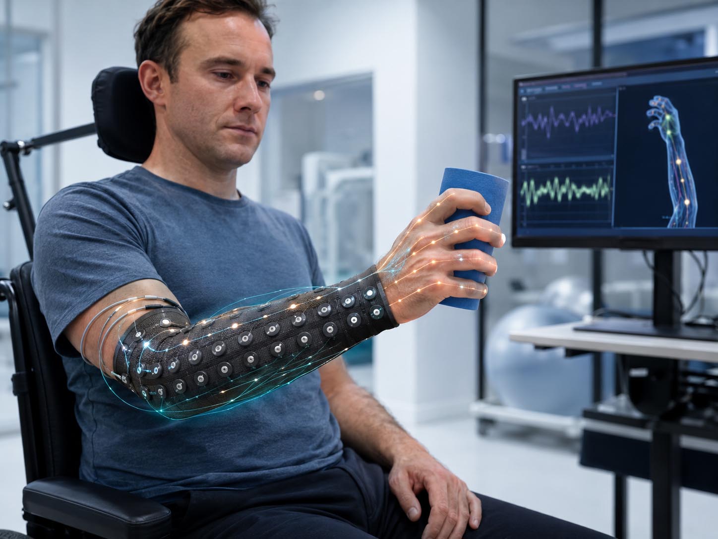
Electrical stimulation plus intensive training improved trunk control in five children with cervical SCI

A small retrospective case series from a paediatric SCI programme found that all five children gained trunk control, and four of five showed arm or hand gains, after around 54 sessions combining neuromuscular electrical stimulation with activity-based training. There was no control group.

Read More

Deep Dive

Paediatric spinal cord injury research is thin on the ground. There are far fewer children with SCI than adults, protocols developed for adult bodies don't transfer cleanly to growing ones, and trials are correspondingly hard to run. So most of what exists is small — and this study, published in the journal *Children* on 12 September 2026, is no exception.

What makes it worth reading anyway is that it looks at the upper body, where the evidence gap is widest.

What was done

The team reviewed records from five children, aged 4 to 15, all with chronic cervical-level spinal cord injury — chronic meaning at least six months post-injury, so past the window where spontaneous recovery does most of its work.

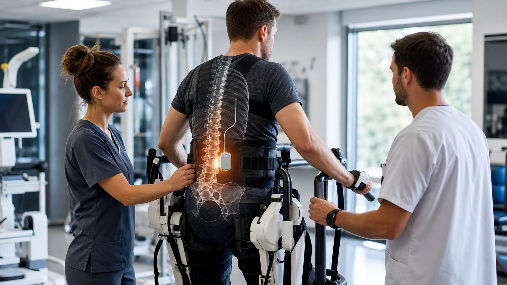
Each child completed an individualised activity-based training programme. Activity-based therapy is built on a straightforward premise: the nervous system below an injury is not simply switched off, and repeated, task-specific practice of real movements — reaching, grasping, weight-bearing, trunk control — can drive it to reorganise. Sessions are intensive and repetitive by design.

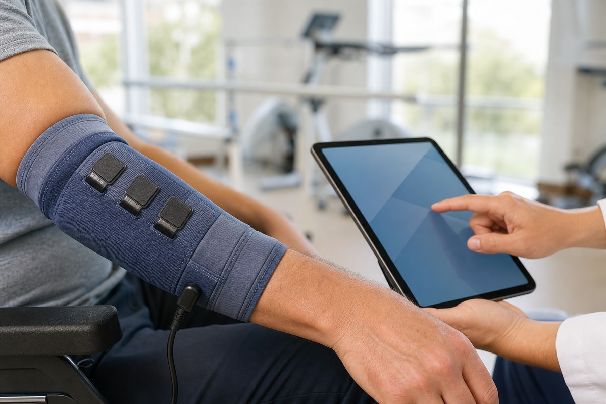
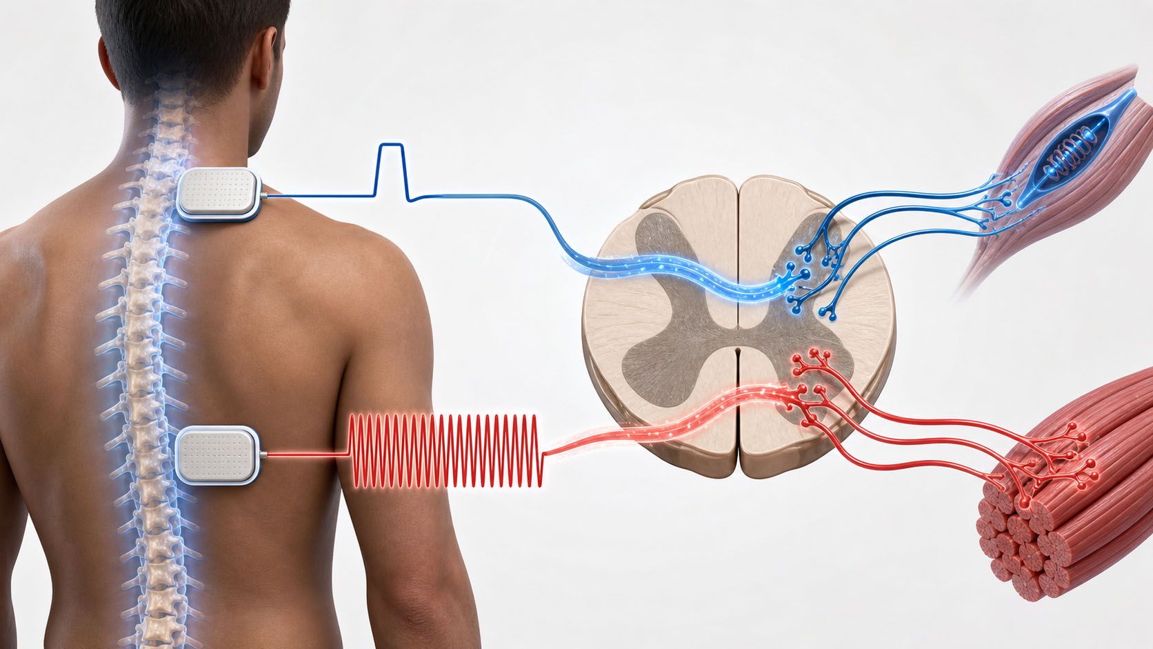
Layered on top of that was wide pulse neuromuscular electrical stimulation (WPS-NMES) — surface electrodes delivering current to make muscles contract during the activity, using longer-than-usual electrical pulses. The "wide pulse" detail matters: longer pulses are thought to recruit sensory nerve fibres more effectively, not just drive the muscle directly, which in principle sends more information back up into the nervous system during the exercise.

Stimulation was delivered with a multichannel device (Xcite, Restorative Therapies) and the settings were tuned per child by the treating therapist. Children completed a mean of 54 sessions.

What changed

Trunk control improved in all five children. Scores on the Segmental Assessment of Trunk Control — a measure that works down the spine segment by segment to find where a child loses the ability to hold themselves upright — rose by between 1 and 6 points.

On the paediatric Neuromuscular Recovery Scale, four of five children gained, mostly on items involving overhead reach and bringing an object to the mouth — the kind of movement that translates directly into self-feeding.

On the Box and Blocks Test, which counts how many blocks a child can move one at a time in a minute, four of five improved in at least one arm. One child stayed at zero in both hands. One child declined in one hand. One child improved at the shoulder and trunk without any change in finger dexterity.

No unanticipated adverse events were reported.

How much weight this can carry

Not much on its own, and the authors are careful about this. Five children, no control group, retrospective review of clinical records rather than a designed trial, and outcomes summarised case by case rather than analysed statistically. The children were also receiving a full multimodal rehabilitation programme, so there is no way to separate what the electrical stimulation contributed from what the training contributed.

The authors' own framing is deliberately modest: improvements were *observed during participation in* the programme, and the findings may help design future controlled studies. That is the right level of claim for this evidence.

Where it has real value is in the pattern of results. Trunk control moved in every child, which is notable given trunk stability is often the thing that unlocks arm function — you cannot reach overhead if you cannot stay upright. And the gains clustered around specific, functional movements rather than appearing everywhere at once.

Reader Q&A

Is this the same thing as FES? Closely related. Functional electrical stimulation (FES) usually refers to stimulation timed to produce a useful movement, such as cycling or grasping. NMES is the broader term for using electrical current to make muscles contract. The "wide pulse" variation used here is a specific parameter choice aimed at recruiting sensory as well as motor fibres.

Why does it matter that these were children? Because almost all of the upper-limb NMES evidence comes from adults, and children are not small adults. Growing musculoskeletal systems, different injury mechanisms, and a developing nervous system all change the calculation. A four-year-old and a fifteen-year-old in the same study also illustrates how wide that range is.

One child got worse in one hand. What does that mean? Honestly, with five children and no control group, it is impossible to say whether that was related to the intervention, to measurement variability, or to something else entirely. It is reported here because the authors reported it, and a study that only listed the improvements would be less trustworthy.

Should families be seeking this out? This study doesn't establish that. It describes what happened in one clinical programme. Activity-based therapy with NMES is available at some specialist paediatric centres, and the safety signal here was reassuring, but the evidence for benefit remains preliminary.

Read full article → (external link)
Research Rehabilitation 17 SEPT 2026

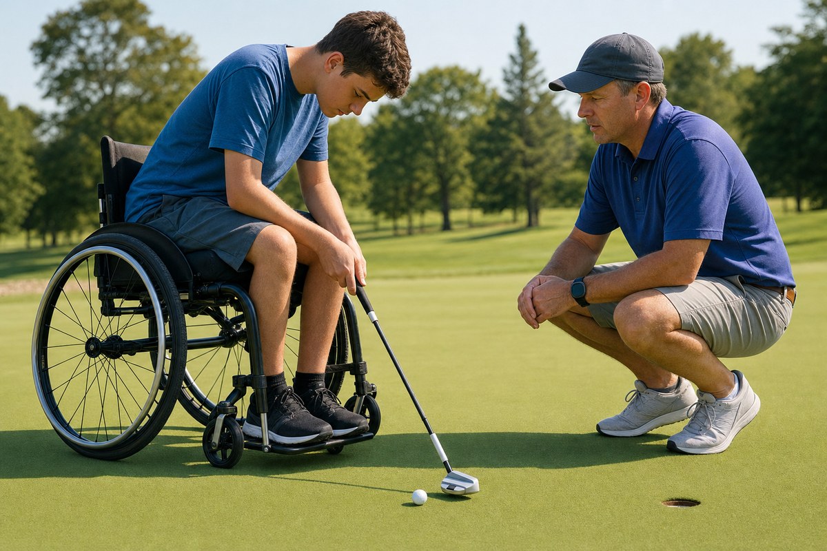
"That's not for me": what public health messages about sitting sound like from a wheelchair

People with SCI sit for 10 to 13 hours a day. Interviews with 12 wheelchair users found they understand the health risks perfectly well — and that the standard 'get up and move' messaging reads as written for somebody else entirely.

Read More

Deep Dive

"Sitting is the new smoking" has been one of the more successful public health slogans of the last decade. A study published in *Healthcare* on 17 September 2026 asked a question that campaign never did: what does that message land like if you use a wheelchair?

The short answer, from the people interviewed, is that it lands as not being addressed to them at all.

The study

Researchers led by Kathleen Martin Ginis — whose group has done much of the foundational work on physical activity after spinal cord injury — ran semi-structured interviews with 12 people. All were medically stable, living in the community, at least a year past injury (average 17.3 years), average age 49, and all used a wheelchair outside the home.

The analysis method was reflexive thematic analysis — a qualitative approach where researchers work through interview transcripts repeatedly to build themes, treating their own interpretation as part of the process rather than pretending to be a neutral instrument. It is designed to surface meaning rather than count occurrences, which is the right tool when the question is "what does this mean to you" rather than "how often does this happen".

For context: people with SCI accumulate an average of 10 to 13 hours of sedentary behaviour per day, well above general population figures.

The five themes

Understanding is built socially, not from health campaigns. Participants had assembled their knowledge of sedentary behaviour from their own experience and from conversations with others in their lives, rather than from public health information.

Sitting worsens symptoms, and symptoms force more sitting. This was described as a loop rather than a one-way effect. Long periods seated aggravate pain, pressure, spasticity and stiffness — and when those symptoms flare, being up and active becomes harder, so seated time increases.

Activity is planned; breaks from sitting are accidental. Participants deliberately scheduled exercise. But the interruptions to sitting that public health guidance emphasises — standing up, shifting, moving around every half hour — happened incidentally, if at all, rather than as an intentional practice.

"Life as a sitter": sitting becomes identity. For people who have used a wheelchair for nearly two decades on average, sitting isn't a behaviour that can be neatly separated out and modified. It is how they exist in the world.

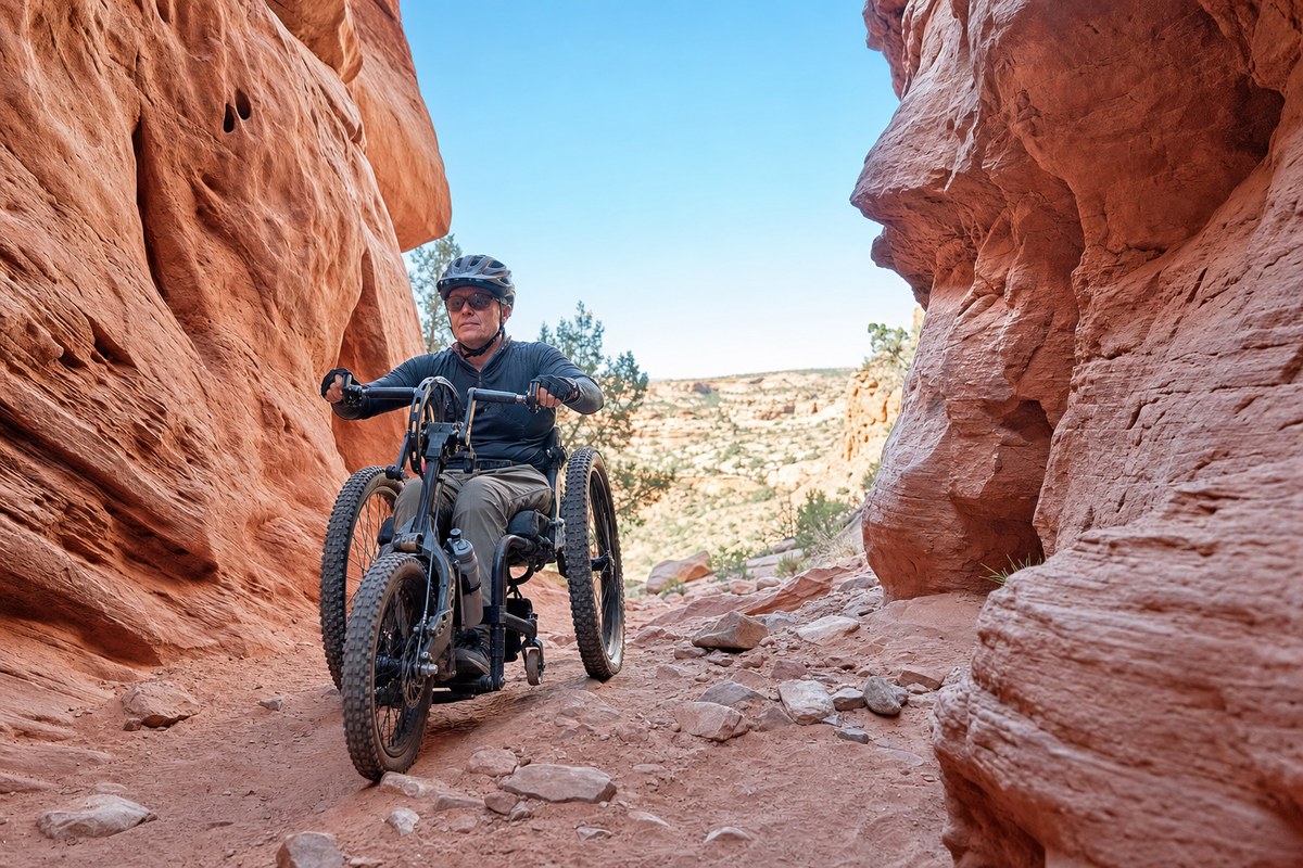
Media messaging is ableist and excludes wheelchair users. This was the sharpest theme. Campaigns built around "stand up", "take the stairs" and "get out of your chair" don't merely fail to apply — they actively signal that the message was written for a body other than yours.

The complication for anyone designing an intervention

The finding that should give programme designers pause is this: participants described sitting as unavoidable, but also as restorative and necessary.

That is not denial or rationalisation. After a spinal cord injury, seated rest genuinely serves functions it doesn't serve for an ambulatory person — managing fatigue, managing pain, recovering from the physical cost of transfers and mobility. An intervention that frames all sedentary time as a problem to be minimised is asking people to give up something they need.

Which means the useful question is probably not "how do we reduce sitting time" but "which sitting, and how is it broken up". Pressure relief practice, for instance, already exists as a routine that interrupts seated time for a completely different reason — and it is exactly the kind of intentional interruption the participants said was missing.

The limits

Twelve people, all community-dwelling, all long post-injury, all wheelchair users, recruited into a single qualitative study. This tells you about the meanings those twelve people brought to the topic. It does not tell you the prevalence of any view, and it says nothing about what would actually change behaviour or health outcomes.

Qualitative work of this kind is best understood as the stage that comes before intervention design — it establishes what the target of an intervention should be, so that the trial that follows isn't built on an assumption nobody checked.

Reader Q&A

Is sitting actually bad for people with SCI, or is this about ambulatory people? The cardiometabolic risks associated with prolonged sedentary time — cardiovascular disease, insulin resistance — are well documented after SCI, and there is a separate body of work on interventions targeting them. What this study addresses is how the messaging around those risks is received, not whether the risks are real.

What would better messaging look like? The study doesn't prescribe one, but the participants' comments point in an obvious direction: messages built around movement rather than standing, that acknowledge seated rest has a purpose, and that offer interruptions a wheelchair user can actually perform.

Why interview only 12 people? Reflexive thematic analysis aims for depth over breadth. The goal is rich accounts from a group with relevant experience, not a representative sample. Twelve is within the normal range for this method — though it does mean the findings shouldn't be read as "people with SCI think X".

Does this mean I should be sitting less? That is a question for you and your clinical team, and the honest answer from this study is that it wasn't designed to tell you. What it does suggest is that the version of the advice aimed at the general public probably needs translating before it is any use to you.

Read full article → (external link)